
The degree of stenosis, the presence or absence of collateral vessels, chronicity of carotid artery disease, its bilaterality, and associated systemic vascular diseases help contextualize the severity of OIS. Bilateral involvement may occur in up to 22% of cases. Men are affected twice as often as women likely due to the higher incidence of atherosclerotic disease in males. Ocular ischemic syndrome occurs at a mean age of 65 years, is rare before 50, has no racial predilection. OIS has important systemic implications, as disease of the common (CCA) or internal (ICA) carotid arteries may cause ipsilateral ocular signs and symptoms that in turn could herald a cerebral infarction. OIS commonly occurs in the elderly with men more affected than women, owing to the higher incidence of atherosclerosis and carotid artery disease in these patients. Principal symptoms include visual loss, transient visual loss, and ischemic ocular pain. Ocular ischemic syndrome (OIS) is a rare, but vision-threatening condition associated with severe carotid artery occlusive disease (stenosis or occlusion) leading to ocular hypoperfusion. 11.3 Extracranial–Intracranial (EC-IC) Arterial Bypass Surgery.10 Management of Ocular Ischemic Syndrome.


The balloon is then deflated and removed, leaving the stent in place. The balloon is inflated, compressing the stent against the plaque in the artery walls and opening blood flow back up. A wire with a tiny balloon on the end is also pushed through the catheter to the blockage site, along with a wire mesh stent. Using the fluoroscopy as a guide, the doctor then threads a tiny catheter through the artery all the way up to the blockage in the neck. “Then the computer subtracts the ‘before’ images from the ‘after’ images, so we get a very clear, high-resolution picture of exactly where the dye is going.” “We take X-rays before and after the contrast dye is injected,” Dr. The doctor uses X-ray technology, called fluoroscopy, to view moving pictures of the arteries through the body. A tiny cut is made in the groin, and a catheter is advanced through the groin artery to the neck, and contrast dye is injected through it. Patients are given local anesthesia at the incision site, and a sedative to relax them. This procedure is done by a vascular specialist. Before angiography or CT scans are performed, patients have contrast dye injected into the bloodstream so that their arteries show up clearly on the images. Usually, it provides a clear enough picture to identify a blockage and determine a treatment plan.īut occasionally, other scans such as magnetic resonance imaging (MRI), carotid angiography (a special type of X-ray) or computerized tomography (CT) are also needed.

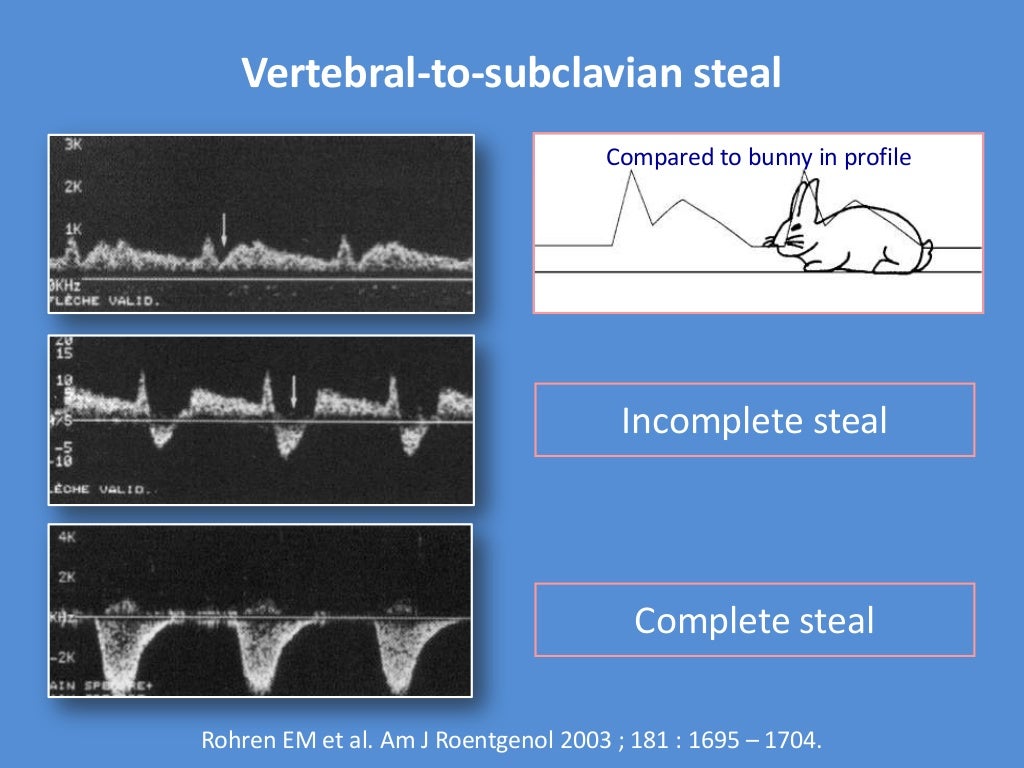
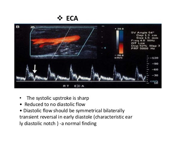
Ultrasound is the preferred diagnostic tool, because it does not expose patients to unnecessary radiation. This is a painless, harmless test that uses sound waves to create an image of the carotid arteries and the plaque inside them. Patients in whom carotid artery disease is suspected are usually tested using a carotid Doppler ultrasound. Dizziness, numbness, weakness or a severe headache may be signs of a TIA caused by a blocked carotid artery. A stroke or a mini-stroke (also called a transient ischemic attack or TIA) can also be a sign that a person’s carotid artery is narrowed or blocked. Carotid artery disease may be suspected if a doctor hears a whooshing sound (called a bruit) while listening to a patient’s neck with a stethoscope during a checkup.


 0 kommentar(er)
